
PET, TOF, and molecular imaging: why time of flight matters
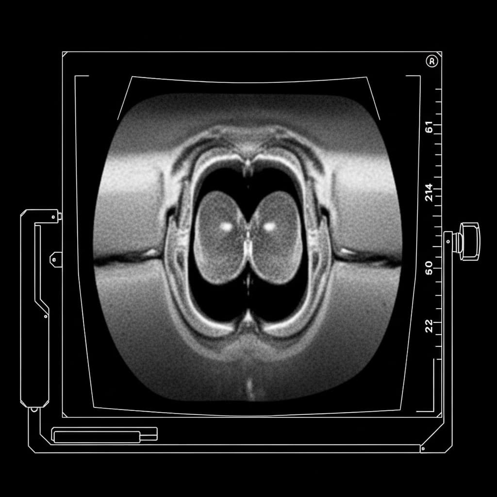
Positron emission tomography (PET) maps the distribution of a radiotracer in the body. Each positron–electron annihilation yields two nearly collinear 511 keV photons; detecting them in coincidence traces a line of response (LOR) between two detectors. Image reconstruction from millions of LORs is the core of the clinical or research system.
When the arrival time difference between the two photons is measured with very high precision (time of flight, TOF), each event adds localization information along the LOR: not only “something happened along this line,” but “with higher probability in this segment.” That reduces noise in the reconstructed image and improves contrast and, in practice, effective resolution for the same acquisition time or dose.
Useful clinical TOF requires fast detectors (crystals, SiPM/SPAD), readout electronics with temporal resolution on the order of hundreds of picoseconds, and a data acquisition (DAQ) system that is not the bottleneck: high count rates, stable synchronization across thousands of channels, and data flow into reconstruction. Detector physics, systems engineering, and acquisition software all meet there.
In molecular imaging and challenges such as early diagnosis, where subtle changes matter, the quality of the PET chain—from detector to algorithm—makes the difference. TOF is not an “extra”: it is a design lever when combined well with the rest of the system.